Digicyte Digital pathology systems with AI support

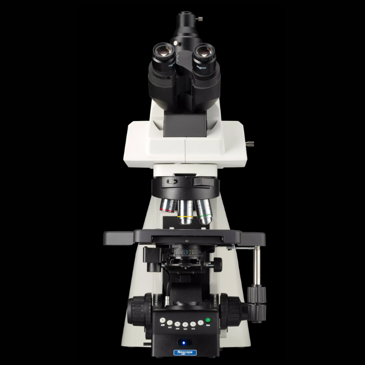
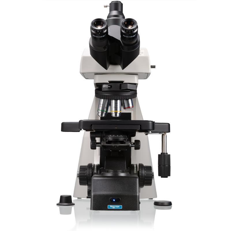
- Nexcope NE900 microscope series systems
- With either a motorized automatic Z-axis and integrated control
- With either a motorized objective changer and motorized stage
- 3W LED illumination with up to 60,000 hours of operating time
- Ergonomic tilting trinocular head (0–35°)
- Digicyte BigEye GS 12MP USB camera
- Digicyte ResoluX Research software
Main Technical Features
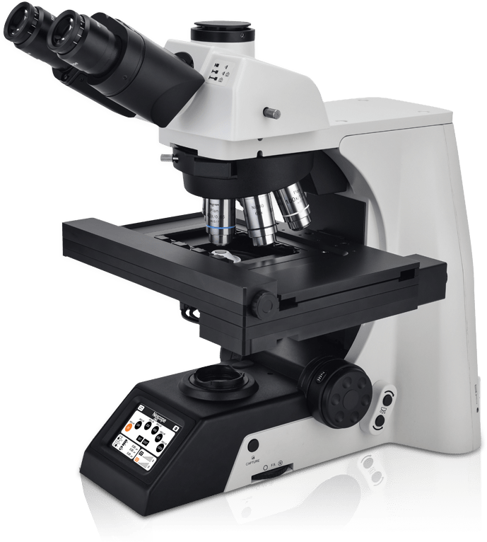
- Flagship NE950 modell with motorized microscope stand with automated Z-axis and integrated controllers
- Full support for motorized objective nosepiece and motorized stage
- 3W LED illumination with up to 60,000 hours lifetime
- Ergonomic tilting trinocular head (0–30°)
- Adjustable interpupillary distance: 47–78 mm
- Light splitting ratios: 100:0 / 20:80 / 0:100
Objective Configuration
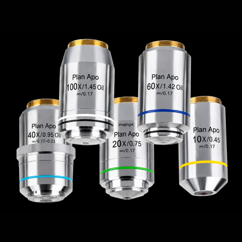
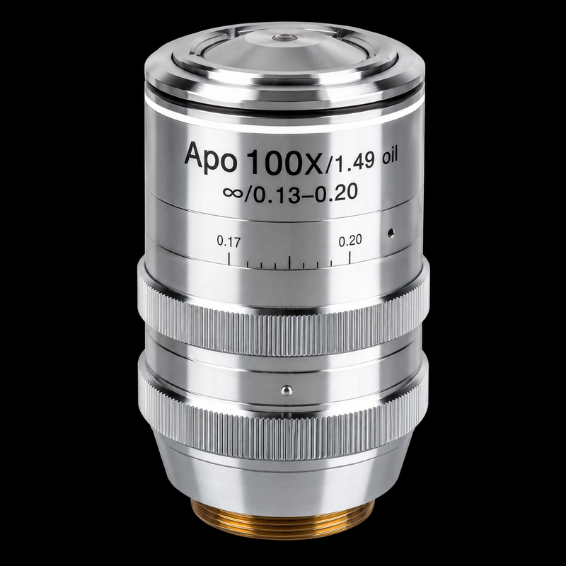
The system can be configured with high-performance semi-apochromatic and apochromatic objectives:
- Semi-Apochromat 4X/0.13
- Semi-Apochromat 10X/0.3
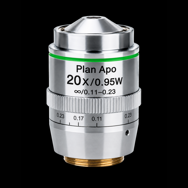
- Apochromat 20X/0.75
- Apochromat 40X/0.95 with correction collar
- Semi-Apochromat 100X/1.4 (optional)
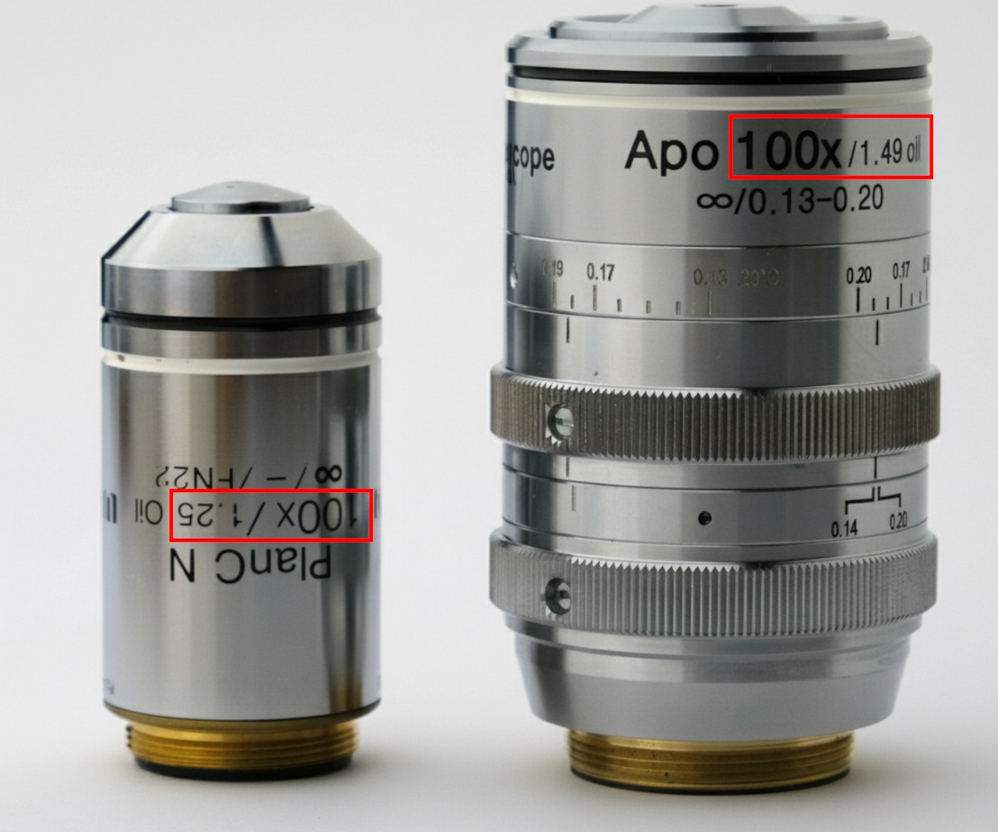
Optional premium objectives are also available depending on the application requirements:
- 100X/1.43 UPlan APO objective
- 100X/1.49 Super UPlan APO objective
Motorized Stage and Batch Scanning
The high-precision motorized X/Y stage features:
- X-axis travel range: 125 mm
- Y-axis travel range: 75 mm
- Positioning repeatability: ±1.5 μm
The system supports automated large-area slide stitching and Batch Scanning of up to four microscope slides simultaneously.
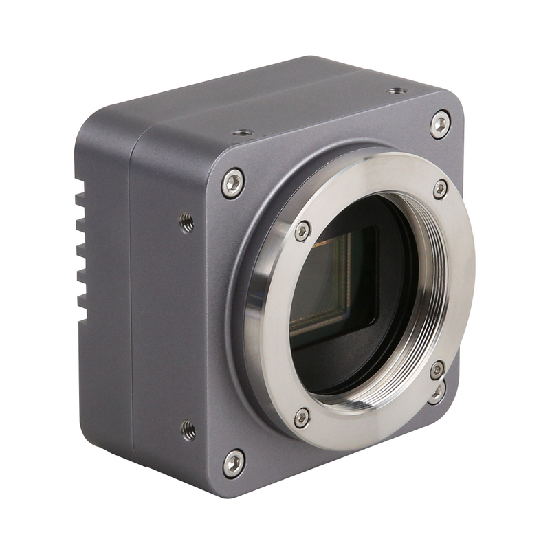
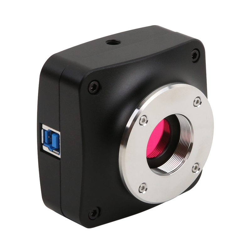
Digicyte BigEye GS 12 MP Camera
- Sony IMX304 Global Shutter CMOS sensor
- 12.3 MP resolution (4096 × 3000 px)
- 23.4 fps at full resolution
- USB 3.0 interface
- 3.45 μm pixel size
- Global shutter technology for distortion-free imaging
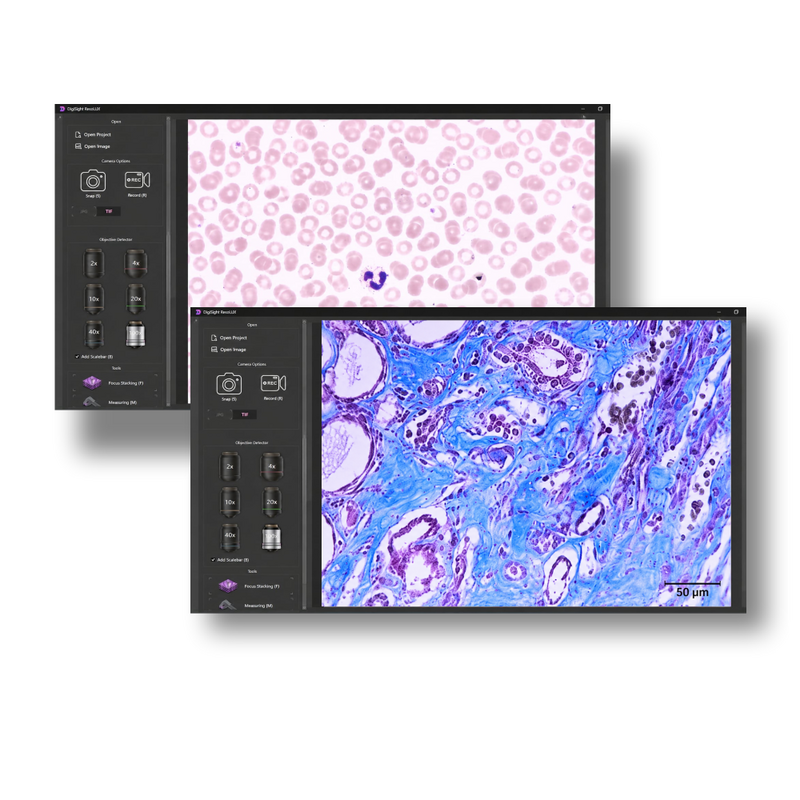
ResoluX AI Image Processing Software
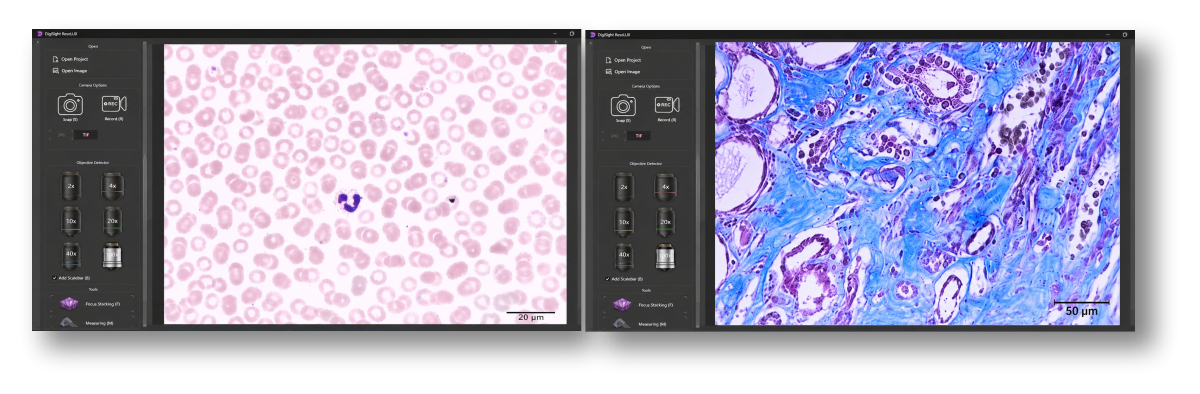
The Digicyte ResoluX platform provides advanced automation and AI-assisted image processing capabilities:
- Automated large-area scanning
- GPU-accelerated EDF / focus stacking
- AI-based Super-Resolution processing
- Real-time chromatic aberration correction
- Vignetting and geometric distortion correction
- “AI True Color” image reconstruction
The Super-Resolution algorithm can increase effective image resolution up to 2× beyond the optical resolution limit of the objective.
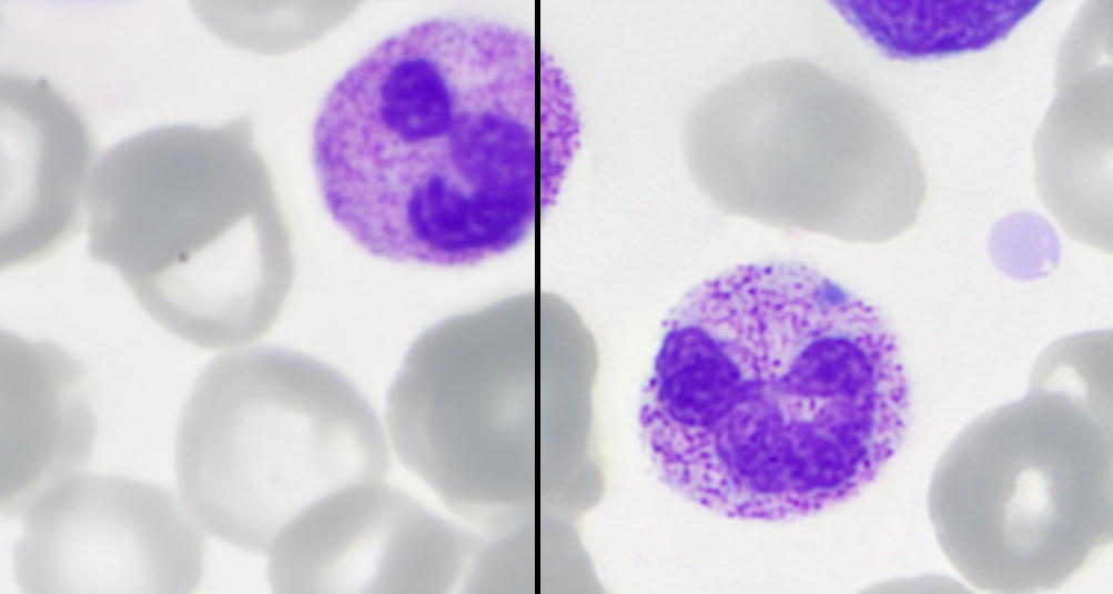
Achieving Up to Twice the Resolution in Brightfield Microscopy
Digicyte.ai is the first — and currently the only — company to successfully implement Sparse Deconvolution technology in brightfield microscopy, enabling image resolution approaching up to twice that typically achievable with conventional 100x oil immersion objectives.
This breakthrough opens new possibilities for both research and clinical applications, particularly in cytology and hematology, where enhanced image detail may support more accurate and confident diagnostics.
Digicyte microscope cameras and the ResoluX software platform are designed to integrate Sparse Deconvolution seamlessly into everyday microscopy workflows.
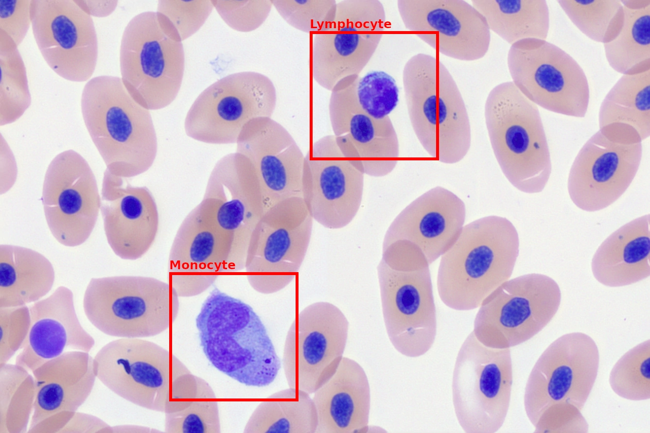
AI-Based Applications for Cell Detection and Classification
Digicyte.ai provides advanced AI-powered solutions for automated cell detection, classification, and segmentation across a wide range of microscopy applications.
With years of experience in developing domain-specific AI models, Digicyte integrates these technologies directly into microscope camera workflows and the ResoluX software platform. Automated cell recognition and measurement capabilities help reduce manual workload while supporting faster, more reproducible image analysis.
The system is suitable for:
- automated cell counting,
- cell size measurement,
- morphological classification,
- object detection,
- and AI-assisted image segmentation.
Numerical Aperture and Resolution
Figure 2 – What Does a Higher Numerical Aperture Mean?
Figure 2 illustrates the concept of numerical aperture. The 10x objective on the left has a relatively low NA and can only collect light from a narrow cone. The 10x objective on the right has a much higher NA, allowing it to capture light from a significantly wider cone.
A wider cone does not simply mean more light — it also means more optical information, including higher-order diffraction patterns from the specimen. This additional information is what enables higher image resolution.
Higher NA = Higher Resolving Power
Figure 3 compares:
- a standard 10X/0.25 objective,
-
and a higher-performance apochromatic 10X/0.40 objective.
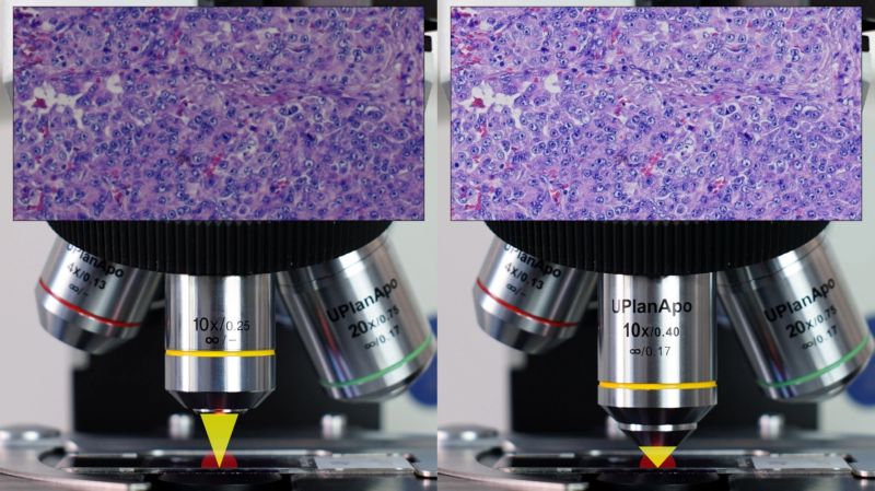
The higher NA objective produces a visibly sharper image and resolves fine structural details that remain blurred with the lower NA objective.
The same principle is demonstrated even more dramatically in Figure 4:
- left: 20X/0.40 objective,
- right: 20X/0.75 objective.
The higher NA objective reveals significantly more detail and image clarity.
The relationship between resolution and numerical aperture is approximately linear:
where:
- = resolution,
- = imaging wavelength,
- = numerical aperture.
This means that doubling the numerical aperture approximately doubles the resolving power of the optical system.
Aperture Is Important Beyond Microscopy
The same optical principles apply in astronomy. Larger telescope apertures collect more light and more information, resulting in higher resolution.
A good example is the comparison between:
- the Hubble Space Telescope with its 2.4 m mirror,
- and the James Webb Space Telescope with its 6.5 m primary mirror.
The significantly larger aperture of JWST enables much higher image resolution, similarly to how high-NA microscope objectives reveal finer microscopic detail.
Why Is Resolution Physically Limited?
Light behaves as a wave. Because of this, microscope objectives cannot focus light into a mathematically perfect point. Instead, they generate an interference pattern called an Airy disk.
This pattern consists of alternating bright and dark rings caused by constructive and destructive interference. Higher numerical apertures produce smaller Airy disks, allowing finer structures to be distinguished from one another.
This physical principle forms the foundation of modern high-resolution microscopy and AI-assisted Super-Resolution imaging technologies.
Airy Disks, Interference, and Resolution
Figure 6 – How a Lens Forms an Image
Figure 6 illustrates how a lens forms an image through interference. When light waves arrive in phase, they reinforce each other and create bright regions (constructive interference). When waves arrive out of phase by half a cycle, they cancel each other out, producing dark regions (destructive interference).
The resulting pattern of bright and dark areas at the focal plane forms the final interference image generated by the optical system.
Figure 7 – The Relationship Between NA, Airy Disks, and Resolution
Figure 7 demonstrates the relationship between numerical aperture, Airy disks, and image resolution.
The same cluster of point-like objects is imaged using objectives with three different numerical apertures:
-
Left – Low NA:
The Airy disks are large and strongly overlap, causing the points to blur together into a single unresolved structure. -
Middle – Medium NA:
Increasing the NA reduces the size of the Airy disks, allowing individual points to become partially distinguishable. -
Right – High NA:
With high numerical aperture, the Airy disks become much smaller and sharper. Individual point sources are clearly resolved as separate structures, revealing significantly more detail.
Why Numerical Aperture Is So Important
Every point source in a specimen produces a characteristic interference pattern known as an Airy disk. The size and sharpness of this pattern are primarily determined by:
- the numerical aperture,
- and the wavelength of light.
Objectives with higher numerical apertures:
- generate smaller and sharper Airy disks,
- capture more diffraction information,
- and therefore provide substantially higher image resolution.
This principle forms one of the fundamental optical foundations of modern high-resolution microscopy, digital image reconstruction, and AI-assisted Super-Resolution imaging technologies.